人的頭顱從外觀上看不到骨頭內的變化,往往從解剖圖裡去預做一個圖型的概念,對進入x光片的解剖相對位置較有助益,,以下請點入![]() 頭骨的縱面解剖圖 先去認識頭骨的剖面圖,從而可以再進入側位x光的圖像做一良好的對照!!
頭骨的縱面解剖圖 先去認識頭骨的剖面圖,從而可以再進入側位x光的圖像做一良好的對照!!
再來就是別忘了,也順便對頭骨的內面那些凹凹凸凸的構造也了解一下,各位一定要了解骨頭那麼硬,怎麼會有痕跡?,想想,別忘了,還有這一顆~~大腦與小腦,,,,腦部的結構會產生壓痕,這就是那些凹凹凸凸的解剖構造, ![]() 頭骨的內部解剖圖 ,有了以上的概念,相信對x光透視所形成的圖像您一定會有更好的影像解釋,原來頭骨是這麼奧妙的呀!再來就是要去了解一下這裡頭的孔的解剖名稱,,,,
頭骨的內部解剖圖 ,有了以上的概念,相信對x光透視所形成的圖像您一定會有更好的影像解釋,原來頭骨是這麼奧妙的呀!再來就是要去了解一下這裡頭的孔的解剖名稱,,,,![]() 頭骨內部的孔的名稱順便也看一下真的骨頭
頭骨內部的孔的名稱順便也看一下真的骨頭 ![]() 真的頭骨內部
真的頭骨內部
當然也要把重要的下顎骨的解剖位置也復習一下→![]() 下顎骨 順便也看一下真的下顎骨 →
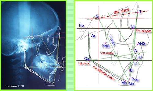
下顎骨 順便也看一下真的下顎骨 →![]() 真的下顎骨有了概念後,就可以直接進入側位片的圖型判讀的領域裡,底下就是一張側位片的圖片,是以電腦繪圖的方式去描繪出來的,這是以醋酸紙(ACETATE PAPER)將X光片疊在上頭,放在燈箱下去以HB鉛筆去描繪的,當然你必須有鉛筆,橡皮擦,量尺,角度尺,這樣才可以測量角度,畫平面,畫角度線,畫牙齒圖形,顎骨與顏面各解剖點的圖,脊椎的骨架,,,,等,得到的就是下列第二張圖的情形,當然必需把各個角度與平面的資料另以紙張記錄下來,得到的是下列的第三張圖所示,這就是測量值,以此就可以進行診斷與治療計畫的擬定,顏面的突度,暴不暴牙,支抗設計,要不要手術?該不該拔牙?需不需要用到骨釘?裝置的使用?這就是這張x光片經過判讀後所得到的這麼多價值,所以矯正治療必需拍這樣的x光片,這樣您瞭了嗎?~~~~~~~~~
真的下顎骨有了概念後,就可以直接進入側位片的圖型判讀的領域裡,底下就是一張側位片的圖片,是以電腦繪圖的方式去描繪出來的,這是以醋酸紙(ACETATE PAPER)將X光片疊在上頭,放在燈箱下去以HB鉛筆去描繪的,當然你必須有鉛筆,橡皮擦,量尺,角度尺,這樣才可以測量角度,畫平面,畫角度線,畫牙齒圖形,顎骨與顏面各解剖點的圖,脊椎的骨架,,,,等,得到的就是下列第二張圖的情形,當然必需把各個角度與平面的資料另以紙張記錄下來,得到的是下列的第三張圖所示,這就是測量值,以此就可以進行診斷與治療計畫的擬定,顏面的突度,暴不暴牙,支抗設計,要不要手術?該不該拔牙?需不需要用到骨釘?裝置的使用?這就是這張x光片經過判讀後所得到的這麼多價值,所以矯正治療必需拍這樣的x光片,這樣您瞭了嗎?~~~~~~~~~

S Sella: Mid point of sella turcica(蝶鞍)
N Nasion: Most anterior point on fronto-nasal suture
Or Orbitale: Most inferior anterior point on margin of orbit(眶下點)
Po Porion: Upper most point on bony external auditory meatus(外耳道孔最上點)
ANS Anterior Nasal Spine(前鼻棘)
PNS Posterior Nasal Spine(後鼻棘)
Go Gonion: Most posterior inferior point on angle of mandible(下顎骨的最後下點)
Me Menton: Lower most point on the mandibular symphysis(下顎骨聯合的最下點)
A point: Position of deepest concavity on anterior profile of maxilla
B point: Position of deepest concavity on anterior profile of mandibular symphysis
 這一個紅色記號處就是gonion
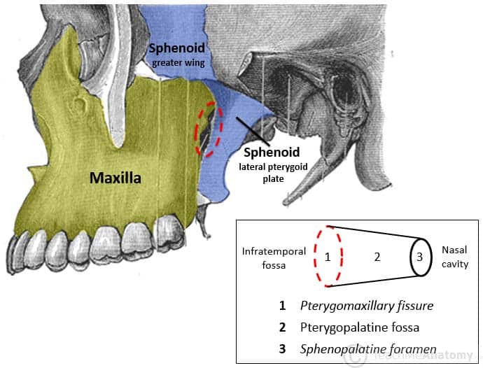
x光裡的Ptm(pterygomaxillary fissure翼狀上頜突縫點)那一個淚滴狀(tear drops)的形狀,在實際頭顱的位置是這樣的→ Ptm ,從全景片裡也可以看到是長這樣的→ Ptm在全景片
Frankfort Plane Po - Or Equivalent to the true horizontal when patient is standing upright.
Maxillary Plane PNS - ANS Gives inclination of maxilla relative to other lines/planes.
Mandibular Plan Go - Me Gives inclination of mandible relative to other lines/planes.
The angle MMPA - Maxilla to Mandibular Planes Angle (Maxillary plane to Mandibular plane) Gives an inclination of the maxilla relative to the mandible, this in turn indicates the relative proportions of face height and acts as an indicator for future growth direction. (MMPA就是上下顎平面的延長線交角,這個角度顯示出顏面的相對高度比例,同時也扮演著將來生長發育的方向)
S - N Line: Indicates orientation of anterior cranial base.(代表前顱底)
N - A indicates relative position of maxilla the cranial base
N - B indicates relative position of maxilla the cranial base
The angles SNA; SNB; ANB indicates relative position of maxilla/mandible to each other and to the cranial base
SNA、SNB角代表上、下颌相對前顱底的前後位置關係,但它們明顯地受前臚底平面倾斜度的影響
Long axis of upper central incisor/lower central incisor (root apex to incisal edge) - allows measurement of the angulation of incisors to maxilla/mandibular planes.
面角(facial angle):指眼耳平面(FH)與面平面(NPg)之間的夾角。反映頦部與面部其他部分的相對位置關係。
下頜平面角(SN-MP):前顱底平面(SN)與下頜平面(MP)之間的夾角。反映下頜平面的陡度及面部的高度。
N-Me:硬組織全面高 (full facial height)
N-ANS:硬組織上面高(upper facial height)
NAP~~代表著是顏面的突度角( facial convexity angles)
ANB顯示出上顎骨與下顎骨的相對位置關係, 可以提供顎骨的大小與位置程度的差異性的測量
以下是白人(高加索種)的ANB測量標準值
ANB 2-4o= Class I skeletal pattern(代表骨性一類關係)
ANB > 4o= Class II skeletal pattern(代表骨性二類關係)也就是暴牙的類型
ANB < 2o= Class Ill skeletal pattern(代表骨性三類關係)也就是戽斗類型
男、女童三級異常咬合之顱底尺寸,大部份區域皆小於正常咬合,在顱底形態的差異上,男童主要發生在蝶鞍周圍,女童則發生在顱底點與下顎關節突交切點中間區
 這就是 ANB角
這是測量顏面垂直高度(facial vertical height)
 AFP代表面後高度(Ar point to the mandibular plane),AFA代表面前高度(the Me point and its ortogonal projection over the palatine plane)
occipital(枕骨)sphenoid(蝶骨)ethmoid(篩狀骨)frontal(額骨)nasal(鼻骨)
 矯正裡的4大平面(S-N/F.H/O.P/M.P)
 著名的TWEED TRIANGLE(TWEED 三角)(FMA,FMIA,IMPA) 1)FMIA = Angle between Frankfurt Horizontal and long axis of lower incisor. (FH平面與下顎門牙的長軸垂線交角)2)IMPA = Angle between long axis of lower incisor and mandibular Plane. (下顎門牙長軸垂線與下顎平面的交角)3)FMA = Angíe between Frankfurt Horizontal and Mandibular Plane. (FH 平面與下顎平面的交角)
 這個就是著名的WITS
 The "Wits" Measurement: the distance along the occlusal plane bctween perpendiculars raised from Points "A" and "B". (A.B.點分別向咬合平面做出的垂直線,其所產生的差別距離就是WITS的測量值)您也常會聽到所謂高角,低角,正常角的案例,東方人的測量值約是以下這樣的正常垂直骨面型1)SN-MP角平均34.3°(±5°),FH-MP角平均27.2°(±4.7°)。2)SN-MP角大于40°,或FH-MP角大于32°,为垂直發育過度,稱為“高角”病例3)SN-MP角小于29° ,或FH-MP角小于22°,反映垂直發育不足,是為“低角”病例。 在矯正拔牙問题上,高角病例和低角病例有不同的考慮:高角病例拔牙標准可以適當放宽,低角病例拔牙要從嚴掌握。這是因為:
⑴高角病例颏部多后缩,治療結束時切牙宜直立一些,以维持協調的鼻-唇-颏關係;較為直立的切牙還可代償骨骼垂直不調,建立適宜的上下切牙之間的形態和功能關係。低角病例情况正好相反,多數患者颏部前突,切牙宜代償性唇倾一些,這樣不僅有利于面形,也有利于切牙的功能。
⑵高角病例咀嚼肌力弱,颌骨骨密度低,支抗牙易于前移、升高,拔牙間隙的關閉比較容易;同时大臼齒的前移有利于高角病例常常伴有的前牙開咬倾向的矯正。低角病例相反,咀嚼力强,骨密度高,支抗牙不易前移、升高,拔牙間隙的關閉主要由前牙遠心移动完成,而前牙過度的内收不利于低角病例常常伴有的前牙深覆合的矯正。
⑶採用推大臼齒向后或擴大牙弓的方法排齊牙列時,可以造成下颌平面角的開大,這對于高角病例的面形和前牙覆合均產生不利的影响,但對于低角病例却較為有利。
在決定拔牙的牙位時高角與低角病例也有差别:高角病例若拔除靠后的牙齒有利于前牙開咬的控制;低角病例若需要拔牙,宜拔除靠牙弓前部的牙齒,这样不僅易于關閉拔牙間隙,且有利于咬合打開。
6. 矢狀骨面型當上下颌牙弓矢狀關係協調、ANB角正常時,如果需要拔牙,通常是上下牙弓同時對稱性拔除(除非Bolten指数不調)。但若存在上下牙弓矢狀關係不調,决定是否拔牙時應考慮上下牙弓之間的差異。II類错咬上颌牙弓相對靠前、下颌牙弓相對靠後,ANB角較大,為代償這種骨骼不調,治療结束時下切牙可以稍唇倾,下颌拔牙應謹慎。III類错合相反,由于上颌相對發育不足、下颌相對過大,ANB角较小,治療结束時允許上切牙稍稍唇倾,下切牙稍稍舌倾,以代償III類骨骼畸形,上颌的拔牙特别慎重。
Angular measurements
l SNA: Evaluate anterior posterior position of the maxilla.range 82 +/- 3 degrees.(代表評估上顎的前後差異)
l SNB: evaluate anterior posterior position of the mandible. Range 80 +/- 3 degrees.(代表評估下顎的前後差異)
l ANB: indicate the amount of skeletal jaw discrepancy.range 2 +/- 2 degrees.(顯示出上下顎顎骨骨頭的差異量)來看出屬於那一類的錯咬合
l S-N-N- Pog: facial angle, used to classify type of the face as mesomorphic or normal ,Retrognathic when the mandible is retruded and prognathic when mandible is protruded.range is 80 +/- 2 degrees.(S-N與N-Pog平面的交角,稱為顏面角,這可以看出顏面是屬於那一種類型,是前凸還是後縮,角度越大,代表下顎越後退,也就是暴牙的傾向居多,角度越小,顯示出下顎越突出,屬於底包天的戽斗臉型)
l Y- axis: growth axis : intersection of S-Gn plane and FH.it used to determine whether the growth of the mandible is in more horizontal or vertical direction.(Y軸所代表的意義就是生長軸向,它是S-Gn平面與FH平面的中分線,可以判斷出下顎骨的生長是較水平或是較垂直的方向)
l FMA: Go-Gn intersect FH , indicate the vertical height of the ramus.(FMA代表的是Go-Gn平面與FH平面的交角,所代表的意義就是下顎ramus的垂直高度)
l Gonial angle: Go-Gn-Ar : range 126 degrees,angle between ramus and body.(Gonial angle是由Go-Gn-Ar三者所構成的角度,角度越大代表下顎越凸,戽斗的情形越大,角度越小,表示上顎發育較發達,顯出暴牙機率較大)
 nasolabial angle(鼻唇角)
Dental analysis
l 1/ - SN :axis of upper central with SN.evaluate anterior posterior inclination of upper central.range 104 +/- 4.(上顎中門牙的長軸垂線與S-N平面的交角,顯示出中門牙的前後軸向)
l 1\ - MP: axis of lower central to MP.range 90 +/- 5.(指下顎的中門牙與M-P平面的交角)
l 1\- A-Pog: measure distance between tip of lower central to A-Pog regardless of inclination.
l 1/ - A-Pog: axis of upper central to A-Pog.
l 1/ - 1\: interincisal angle. Angle between the long axis of upper central and long axis of lower central. Range 135 +/- 5 ,increase with retrusion.(上下顎中門牙的交角,越大角度,門齒越有直立的現象)
l 1/ - NA: measure distance between the incisal edge of upper central and NA in mm.
l 1\- NB: measure the distance between the incisal edge of lower central and NB.
Vertical planes & lines有
l Facial plane. N- Pog(面平面)
l Maxillary depth plane N-A(上顎深度平面)
l Mandibular depth plane N-B(下顎深度平面)
l Axis of max central – most protrusive –apex
l Axis of mand central- most protrusive-apex
l Growth axis. Y axis.S- Gn.(生長軸向)
矯正治療到結束後必需做一項對照的疊影圖(superimosition)也就是如這樣上下顎的疊影圖 從疊影圖的變化量去了解到上下顎的生長量,與顎骨的發育方向的量與改變,因此這個疊影圖是矯正訓練必須要做的,這叫有始有終!有了x光片的判讀後,就可以配合口內口外的圖像,做出診療計畫,如下圖,深咬的案例,就可以著手與病人討論,開始進入評估階段與溝通治療的施行,一個矯正治療就是這樣開始的~~~~~~~~~~ cephalometrics summary
cephalometric tracing的35張slides(圖的右下角有一個幻燈片show的按鈕)
有關於面前高(anterior facial height)的論述
lateral view x ray tracing●Definitions OF Cephalometric Skeletal Landmarks(英文字義解釋) ●經典的測臚x光片教學slides
A CEPHALOMETRIC EVALUATION OF BRAZILIAN PREHISTORIC MAN
Validity and reproducibility of cephalometric measurementsobtained from digital photographs of analogue headfilms顎三角形分析:一種矯正診斷學的輔助工具;CDJ-12(2):P56-70(1993)

請先 登入 以發表留言。